
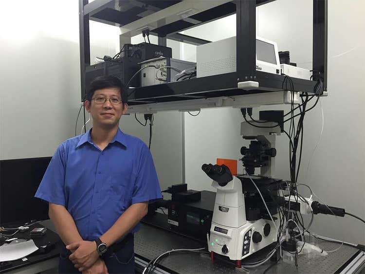
Institute of Atomic and Molecular Sciences uses MetaMorph software to study proteins at the transition zone of primary cilia
COMPANY/UNIVERSITY
Academia Sinica, Institute of Atomic and Molecular Sciences
TEAM MEMBERS
Jung-Chi Liao, PhD, Associate Research Fellow
PRODUCTS USED
MetaMorph Microscopy Automation and Image Analysis Software
The Challenge
The Solution
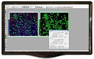
They use MetaMorph® Software with super-resolution image processing to perform two-color fluorescent imaging and have determined the relative positions of proteins at the ciliary base.
MetaMorph Microscopy Automation and Image Analysis Software [Discontinued]
Products Used
MetaMorph® Microscopy Automation & Image Analysis Software is the industry standard for automated microscope acquisition, device control, and image analysis, bringing microscopists greater understanding of cell morphology, function, and behavior for over 25 years. It is the ideal "glue" for easily integrating dissimilar fluorescent microscope hardware and peripherals into a single custom workstation, while providing all the tools needed to perform meaningful analysis of acquired images. The software offers many user-friendly application modules for biology-specific analysis such as cell signaling, cell counting, and protein expression.