
Colorectal Cancer (CRC) Organoids
Patient-derived colorectal cancer organoids
Colorectal cancer patient-derived organoids (PDOs) are three-dimensional (3D) cell culture models that replicate the intestinal epithelial physiology and genetic characteristics of the CRC patient. These CRC PDOs model cell-cell interactions and the influx of oxygen and nutrients within the tumor microenvironment. When cultivated in the laboratory, these tumor organoids serve as multicellular mini replicas of the 3D tumor and have demonstrated the ability to retain their in vivo characteristics.
CRC organoids (and those from other organs in the body) are derived from adult stem cells isolated from patient biopsies. The tissue is processed in the lab and embedded in a gelatinous basement membrane matrix that provides physical support and growth factors. The stem cells from within the tissue divide to form multiple cell types (including more stem cells) that self-assemble into three-dimensional structures mimicking the architectural conformation and physiology of the original tumor. Once formed, these structures can be split apart to re-seed multiple new PDOs that self-assemble in the same way. In this way, the numbers of organoids can be expanded to provide many more, as required.
Advantages of colorectal cancer patient-derived organoids
The representation of CRC-associated genetic alterations and tumor composition make CRC PDOs ideal for cancer research and drug discovery. Biologically relevant CRC PDOs can exhibit drug responses similar to that of patients, pointing to their value in predicting the efficacy of novel therapeutics. However, traditional manual organoid assay workflows do not lend themselves to reproducible high-throughput compound screening for drug discovery.
Advancements in organoid scaling and assay automation have helped to standardize CRC PDO workflows. Our 3D Ready Organoids are manufactured in an industrial environment using our proprietary bioreactor technology and rigorously controlled processes. Many millions of standardized, consistently sized, and quality-tested organoids can now be generated in a single batch. The CRC organoids are cryopreserved and can be used when required for high-throughput assays. Using liquid handling to plate the PDOs in multi-well dishes and treat with compounds significantly reduces errors and hands-on time. Changes upon drug treatment can be evaluated using suitable assays and image analyses.
Colorectal cancer tumoroid culture and assay workflow
The assessment of drug effects on colorectal tumoroids can be automated. The workflow below depicts the automated assessment of anticancer compound effects. PDO scale-up is achieved through expansion in a bioprocess using bioreactor technology. After treating the organoids with the agent to be tested, morphological analysis is performed by ImageXpress® Micro Confocal High-content Imaging System, while SpectraMax® iD5 Multi-mode Microplate Reader is used for the viability assay. The workflow includes automated incubation and liquid handling as well as scheduling software for transferring the plates from the incubator to the liquid handler and to the plate reader.

Experimental protocol for CRC PDOs
- Organoid Bioprocess - CRC PDOs are expanded manually and seeded into bioreactors. The resulting organoids are counted and assessed to determine their final size-range. A defined number of organoids are placed in vials and cryopreserved so that they are assay-ready at the user’s convenience.
- Thaw - PDOs are thawed at 37°C washed with fresh media and pelleted.
- Seed - PDOs are suspended in the required hydrogel or basement membrane matrix for seeding multi-well plates at the required density.
- Treat - 48 hours after seeding, the compounds or agents to be tested can be applied to the PDOs in the wells at the required concentrations.
- Image and Analyze - Transmitted light (TL) imaging is performed five days after compound treatment to determine desired parameters such as the number of organoids, average diameter, average volume, total area covered by organoids, morphology etc.
- Viability Assay – An example of an End-Point Assay protocol
- Viability Assay Reagent - An automated liquid handler is used to add the assay reagent to the wells for a luminescent ATP cell viability assay, while automated microplate shaking mediates cell lysis.
- Incubation - Incubation is performed at room temperature for 25 minutes.
- Luminescent ATP Cell Viability Assay - Scheduling software and a sample handler are used to transfer the microplate from the incubator to the liquid handler and the microplate reader. Results are analyzed with microplate reader control and data analysis software.
We provide extensive lab automation solutions for custom colorectal organoid workflows. Each step—from organoid culture and seeding to liquid handling and image acquisition—can be automated and combined with our data analytics software solutions to generate regulatory-compliant and publication-ready reports on your organoid assays.
Featured asset
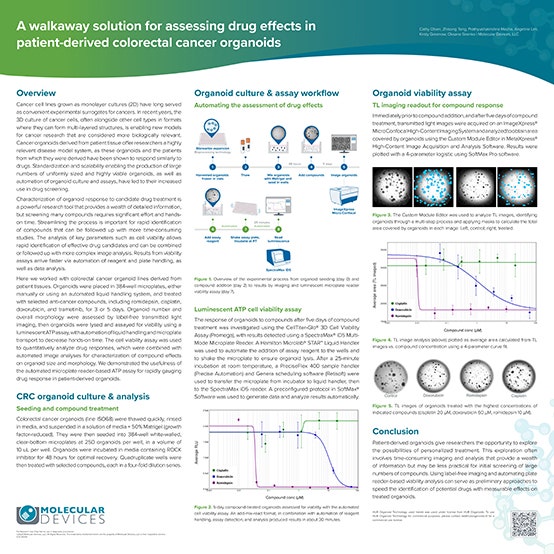
A walkaway solution for assessing drug effects in patient-derived colorectal cancer organoids
In this scientific poster, patient-derived colorectal cancer organoids were cultured and treated with anticancer compounds romidepsin, cisplatin, doxorubicin, and trametinib.
Label-free transmitted light imaging revealed does-dependent changes in the organoid number and the total area occupied by organoids in response to drug treatment. Plate reader-based luminescent ATP assay helped glean quantitative cell viability data from the organoids.
The workflow involves automated liquid handling, microplate transfer, and software analysis methods to decrease hands-on time and accelerate drug screening.
Latest Resources
The complexity of colorectal organoid structures calls for advanced imaging and analysis strategies. Additionally, time consuming and laborious steps must be automated for quicker turnaround and increased consistency. Learn how qualitative and quantitative high-throughput tumoroid analysis can be achieved via automation strategies.