3D Bioprinting
Leverage automated, high-throughput 3D bioprinted cell models for research, drug discovery and toxicology.
What is 3D bioprinting?
Three-dimensional (3D) bioprinting technology is a cutting-edge solution that involves the layer-by-layer deposition of biological materials—including various cell types, bio-inks, and growth factors—to create 3D structures that mimic the architecture and function of 3D cellular models. This process is similar to traditional 3D printing but is specifically tailored for biological applications.
Key 3D bioprinting components include:
- Bioinks: These are the printable materials containing living cells and other biomaterials. They serve as the building blocks for creating 3D cellular models.
- Printer: Specialized 3D printers used in bioprinting allow precise control over the placement of cells and biomaterials.
- Software: Optimal software has the ability to design custom 3D printed structures by programming bioinks into specific patterns. Software should also ensure custom workflows can be stitched with other tasks.
https://vids.moleculardevices.com/watch/Ru4dmoEYsvT6nVceekhhWL
BioAssemblyBot 400 (BAB400) bioprinter seamlessly integrates with ImageXpress high-content screening system for an automated 3D biology workflow.
Optimize bioprinting for 3D biology
Different types of bioprinters for 3D biology use various techniques to print and dispense cells. Some of these include extrusion, inkjet, stereolithography, laser-assisted, and fused-deposition modeling. Most techniques use cell-laden hydrogels, which don’t properly mimic the complex environment needed for organoid development. Whereas extrusion-based printers, such as the BioAssemblyBot® 400, adequately support these environments with the additional benefit of being able to increase scalability in a short amount of time. In addition, extrusion-based printers facilitate the use of a wide array of bioinks, such as cell aggregates, microcarriers, hydrogels with cells, decellularized matrix components, microcarriers, and many more. Extrusion-based bioprinting enables high cell density printing, it’s easy to implement, enables anatomically porous structures, is easy to learn, the hardware is more affordable, and forms biocompatible structures and decreases cell damage.
Potential for Emerging 3D Bioprinting Applications
The advent of 3D bioprinting has ushered in a new era in biomedical engineering, offering transformative solutions across tissue engineering, drug development, disease modeling, personalized medicine, and regenerative therapy. This innovative technology enables the precise fabrication of functional biological structures, revolutionizing research and healthcare with its potential to address critical challenges and enhance therapeutics.
- Tissue Engineering: Bioprinting holds the potential to revolutionize the field of tissue engineering by creating functional miniature organs or “organoids” that can be used to study biological structures. This has the potential to address the shortage of true representative models of in vivo structure and function.
- Drug Testing and Development: Bioprinted models can be used for drug testing to provide more accurate representation of how drugs will behave in the human body compared to traditional cell cultures. This can potentially reduce the need for animal testing and improve the efficiency of drug development.
- Disease Modeling: Bioprinting allows the creation of realistic models that represent human tissues and organs which can provide researchers with a better understanding of diseases and enable the development of targeted treatments.
- Personalized Medicine: The ability to create tissues and organs with a patient's own cells opens the door to personalized medicine. This can lead to treatments tailored to an individual's unique genetic makeup and reduce the risk of potential drug failures.
- Regenerative Medicine: Bioprinting can contribute to regenerative medicine by providing a means to repair or replace damaged tissues and organs. Although their applicability has not been addressed in humans, they have the potential to enhance the body's natural healing processes. They may act as an important bridge between preclinical and clinical studies if protocols can be cleared to enable the vast and direct use of this technology in regenerative medicine.
Bioprinting assay workflow
Our bioprinting platform features an advanced automated enclosure, BioAssemblyBot 400 (BAB 400) for the construction of 3D model systems with increased throughput and precision, eliminating common concerns associated with manual workflows. The BAB 400 utilizes a six-axis robotic arm equipped with interchangeable 'BAB hands' to efficiently produce and maintain living tissues, organoids, and spheroids. It seamlessly integrates with our ImageXpress Confocal HT.ai High-Content Imaging System, equipped with IN Carta Image Analysis Software, a powerful AI/machine learning image analysis software. This turnkey automated solution ensures a fully optimized 3d bioprinting assay workflow.
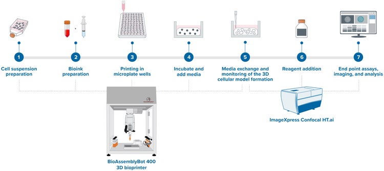
- Cell suspension preparation: Cells are harvested and counted to be resuspended in cell culture medium in required concentration.
- Bioink preparation: Assay-specific extracellular matrix is mixed with cell suspension in necessary concentrations and specific temperatures.
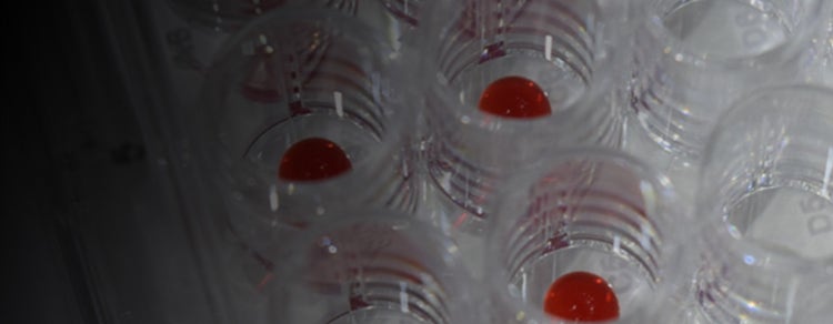
- Printing in microplate wells: The robotic arm’s printhead “hand” prints out the bioink with cells into the cell culture plates.
- Incubate and add media: Printed structures are incubated on the stage or incubator based on temperature and time requirements. The cells in structures are fed with growth factor media specific to the cell line and assay conditions.
- Media exchange and monitoring of the 3D cellular model formation: Cells are fed periodically to replenish the depleted nutrients. These cells in structures are incubated and grown while acclimatizing for further assay steps.
- Reagent addition: Various reagents—for example, growth factors, drugs or even dyes—are added when required in the workflow for growth, drug screening or biomarker-based imaging.
- Endpoint assays, imaging, and analysis: Final steps of the assay workflow are carried out to collect relevant data. The data collected is then analyzed using standard or customized methods.
While bioprinting still has a few challenges to overcome in improving bioink formulations, model vascularization, and cellular model functionality, it is a promising and quickly advancing technology. Despite these challenges, researchers are rapidly bringing bioprinting closer than ever to mimic in vivo structures and functions to deliver targeted, efficient, and effective therapies.